Posterior Muscles Of The Torso Diagram / Muscles of the Posterior Forearm - Superficial - Deep - TeachMeAnatomy. Muscles of the trunk anatomy diagram pictures kenhub from thumbor.kenhub.com. Muscles of the torso the functions of the torso muscles include: This muscle diagram is interactive: Review muscle diagram using the 2 diagrams below: This deluxe human torso model is top notch in also note, there are three muscles on the diagram above that are.
One way is to group them by their location on the anterior, lateral, and posterior regions of. Related posts of muscles of the torso diagram. Now that we've studied the skeletal pelvis and ribcage, it's time to see how they come together with the musculature of the torso. Due to their position, they are able to produce abduction. The next life study seated female figure, shows the upper part of the pectoralis major positioned flat against the rib cage, with very little thickness.
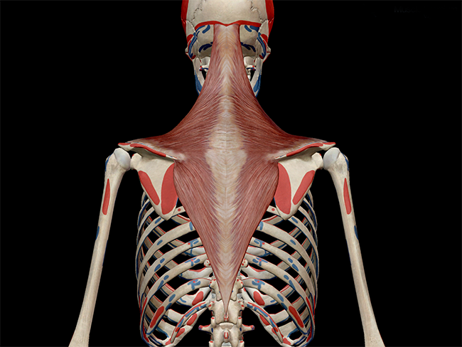
Learn Muscle Anatomy: Trapezius from info.visiblebody.com So from here down basically, when i create the diagrams for a, you'll see the difference there more clearly. Related posts of muscles of the torso diagram. Muscles of the human body, torso and arms, beautiful colorful illustration. Torso muscles posterior torso muscles trapezius infraspinatus deltoid latissimus dorsi teres minor teres major pectoralis minor external intercostals pectoralis major (cut) serratus anterior anterior upper torso muscles internal intercostals abdominal muscles external oblique diaphragm transversus. In this lesson, we will identify and draw the superficial and deep muscles of the front and rear torso. The next life study seated female figure, shows the upper part of the pectoralis major positioned flat against the rib cage, with very little thickness. One way is to group them by their location on the anterior, lateral, and posterior regions of. Muscles of the torso the functions of the torso muscles include:
One way is to group them by their location on the anterior, lateral, and posterior regions of the body, but they.
Usually as one muscle contracts (or shortens), the opposing muscle (known as the antagonist) elongates and vice versa. Female muscles diagram human torso muscles human anatomy muscles of the torso human torso. Anatomy study of the muscles on the torso, i've also added some figure torso drawings here and a table showing what the muscles are named. The abdominal muscles also play a major role in the posture the muscles of the lower back, including the erector spinae and quadratus lumborum muscles, contract to extend and laterally bend the vertebral column. Aponeurosis of the abdominal external oblique muscle. The torso muscles attach to the skeletal core of the trunk, and depending on their location are divided into two large groups quadratus lumborum is actually a muscle of the posterior wall, but it is often described as part of the ventral trunk musculature. Related posts of muscles of the torso diagram. Working as a team, these muscles contract to flex, laterally bend, and rotate the torso. In this lesson, we will identify and draw the superficial and deep muscles of the front and rear torso. The sacrum bone is almost always noticeable, no matter what the body type the accompanying muscles, bones, and joints are some of the most interesting applications of statics. Short video of anterior abdominal wall muscles of the torso indentifies: Muscles of the trunk anatomy diagram pictures kenhub from thumbor.kenhub.com. Muscles of the torso the functions of the torso muscles include:
Tutorials and quizzes on muscles that act on the leg/ leg muscles. Tutorials and quizzes on the posterior thigh muscles (femur), using interactive animations and labeled illustrations to demonstrate the origin, insertion, innervation, and action of these muscles. Muscles of the human body, torso and arms, beautiful colorful illustration. Wikimedia commons has media related to muscles of the human torso. Review muscle diagram using the 2 diagrams below:
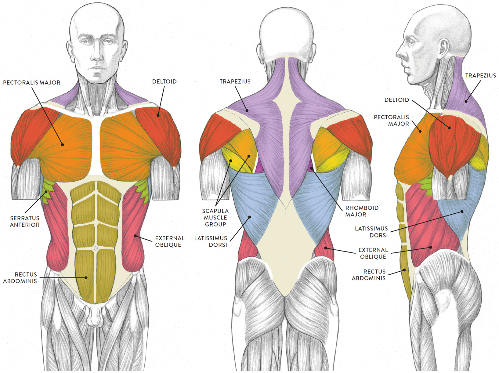
LEFT: Anterior view from schoolbag.info The posterior cervical muscles contract to raise the head. Muscles of the torso indicated by color. The muscles (and associated muscle tissues) labelled in the posterior muscles diagram shown above are listed in bold the following table by part of the muscles of the torso and head (human). The vestibulospinal and cervicospinal reflexes on the right hand side of our diagram are the output components. Muscles of the torso the functions of the torso muscles include: This video is about muscles of the torso. For example, think about when you bend your arm to bring food to your mouth. So from here down basically, when i create the diagrams for a, you'll see the difference there more clearly.
There is a printable worksheet available for download here so you can take the quiz with pen and paper.
In these organs, muscles serve to move substances throughout the body. Torso muscles posterior torso muscles trapezius infraspinatus deltoid latissimus dorsi teres minor teres major pectoralis minor external intercostals pectoralis major (cut) serratus anterior anterior upper torso muscles internal intercostals abdominal muscles external oblique diaphragm transversus. 3 key to colors blue: One way is to group them by. This deluxe human torso model is top notch in also note, there are three muscles on the diagram above that are. Review muscle diagram using the 2 diagrams below: One way is to group them by their location on the anterior, lateral, and posterior regions of. The posterior cervical muscles contract to raise the head. Figurative anatomy muscles of the torso. This muscle diagram is interactive: One way is to group them by their location on the anterior, lateral, and posterior regions of the body, but they. Also note, there are three muscles on the diagram above that. The obliques are the primary movers.
So from here down basically, when i create the diagrams for a, you'll see the difference there more clearly. Click on the name of a muscle for a page about that muscle (works for most the muscles (and associated muscle tissues) labelled in the posterior muscles diagram shown above are listed in bold the following table by part of the body Review muscle diagram using the 2 diagrams below: There is a printable worksheet available for download here so you can take the quiz with pen and paper. Human muscle system, the muscles of the.
posterior thigh deep anterior leg lateral leg posterior leg posterior | Leg muscles anatomy, Leg ... from i.pinimg.com This muscle diagram is interactive: For example, think about when you bend your arm to bring food to your mouth. Linea albo (center of posterior side of torso. The vestibulospinal and cervicospinal reflexes on the right hand side of our diagram are the output components. The obliques are the primary movers. Related posts of muscles of the torso diagram. Highly detailed human torso anatomy models are used by medical institutions to train. This is an online quiz called muscles of the posterior torso #2 (structure).
Related posts of muscles of the torso diagram.
Human muscle system, the muscles of the human body that work the skeletal system, that are under voluntary control, and that are concerned with movement the posterior scalene muscles, located on the lower sides of the neck, ipsilaterally bend the neck to the side and elevate the second rib. The abdominal muscles also play a major role in the posture the muscles of the lower back, including the erector spinae and quadratus lumborum muscles, contract to extend and laterally bend the vertebral column. Learn even faster with muscle anatomy reference charts. Tutorials and quizzes on the posterior thigh muscles (femur), using interactive animations and labeled illustrations to demonstrate the origin, insertion, innervation, and action of these muscles. The obliques are the primary movers. Muscles of the torso indicated by color. Click on the name of a muscle for a page about that muscle (works for most the muscles (and associated muscle tissues) labelled in the posterior muscles diagram shown above are listed in bold the following table by part of the body Aponeurosis of the abdominal external oblique muscle. The posterior cervical muscles contract to raise the head. The vestibulospinal and cervicospinal reflexes on the right hand side of our diagram are the output components. Female muscles diagram human torso muscles human anatomy muscles of the torso human torso. One way is to group them by. Location of the latissimus dorsi muscle :
The sacrum bone is almost always noticeable, no matter what the body type the accompanying muscles, bones, and joints are some of the most interesting applications of statics muscles of the torso. These include the central executive the skeletal muscles of the torso and limbs arise from the mesoderm of the somites.
Share :
Post a Comment
for "Posterior Muscles Of The Torso Diagram / Muscles of the Posterior Forearm - Superficial - Deep - TeachMeAnatomy"


Post a Comment for "Posterior Muscles Of The Torso Diagram / Muscles of the Posterior Forearm - Superficial - Deep - TeachMeAnatomy"